Fig. 1.

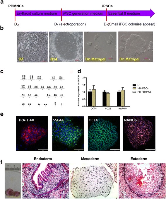
Generation and characterization of patient-specific iPSCs from PBMNCs. a Schematic of patient-specific iPSC generation from PBMNCs. b Small iPSC-like colonies appeared 7 days after electroporation. At 14 days after electroporation, iPSC colonies grew big enough for picking out. After being picked out, iPSCs were cultured on Matrigel-coated plates without feeder. c Karyotype of iPSCs was normal. d qRT-PCR analysis showed expression of OCT4, SOX2, and NANOG of iPSCs (n = 3 independent experiments for each sample). PBMNCs from patient used as negative control, H1 embryonic stem cells used as positive control. e Immunofluorescence staining showed expression of TRA-1-60, SSEA4, OCT4, and NANOG. f Sections of teratomas stained with H&E (endoderm: bronchus; mesoderm: cartilage; ectoderm: epidermis). All scale bars represent 100 μm. PBMNC peripheral blood mononuclear cell, iPSC induced pluripotent cell, D day, GAPDH glyceraldehyde 3-phosphate dehydrogenase, HB hemophilia B
