
Fig. 8.
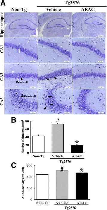
Survival and function in the hippocampus of Tg mice. a After AEAC administration for 4 weeks, brain tissues were collected from subset groups and histological changes were determined as described in the Methods. The slide sections of brain tissue were stained with Nissl and then observed at 400× magnification. b The total number of neuronal cells was calculated per 80 mm2. c AChE activity was measured using the homogenate of hippocampus tissue collected from two mice after AEAC treatment. Data shown are means ± SD (n = 5). #, P < 0.05 compared with the Non - Tg group. *, P < 0.05 compared with the Vehicle treated Tg2576 group