
Fig. 1.
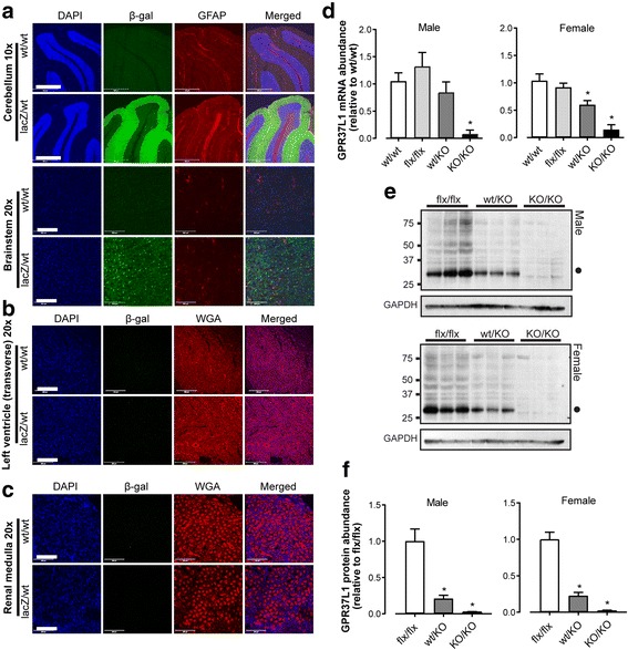
GPR37L1 is abundant in the brain and ablated in EUCOMM-derived knockout mice. a–c Paraformaldehyde-fixed tissue from GPR37L1lacZ/wt and GPR37L1wt/wt mice (mixed-sex, ≥ 12 weeks old) a Brain: nuclei (blue; DAPI); GPR37L1 β-galactosidase reporter (green; β-galactosidase antibody); astrocytes and the Bergmann glia [red, glial fibrillary acidic protein antibody (GFAP)]. Scale bars: 500 μm (× 10 images) and 10 μm (× 20 images). b Heart and c kidney: nuclei (blue, DAPI); GPR37L1 β-galactosidase reporter (green; β-galactosidase antibody); cell membranes [red; wheat germ agglutinin (WGA)]. Images representative of n = 3. Scale bar = 200 μm. d qPCR confirmed loss of cerebellar GPR37L1 mRNA in male and female GPR37L1KO/KO mice (10–12 weeks old, n = 6, one-way ANOVA with Dunnett’s multiple comparisons, *p ≤ 0.05). e Immunoblot and f densitometry of GPR37L1 in cerebellar homogenates from male and female mice (genotypes as indicated, 10–12 weeks old, n = 3, one-way ANOVA with Dunnett’s multiple comparisons, *p ≤ 0.05). Closed circle (Mr ~ 30 kD) indicates predominant, cleaved GPR37L1 species [15]