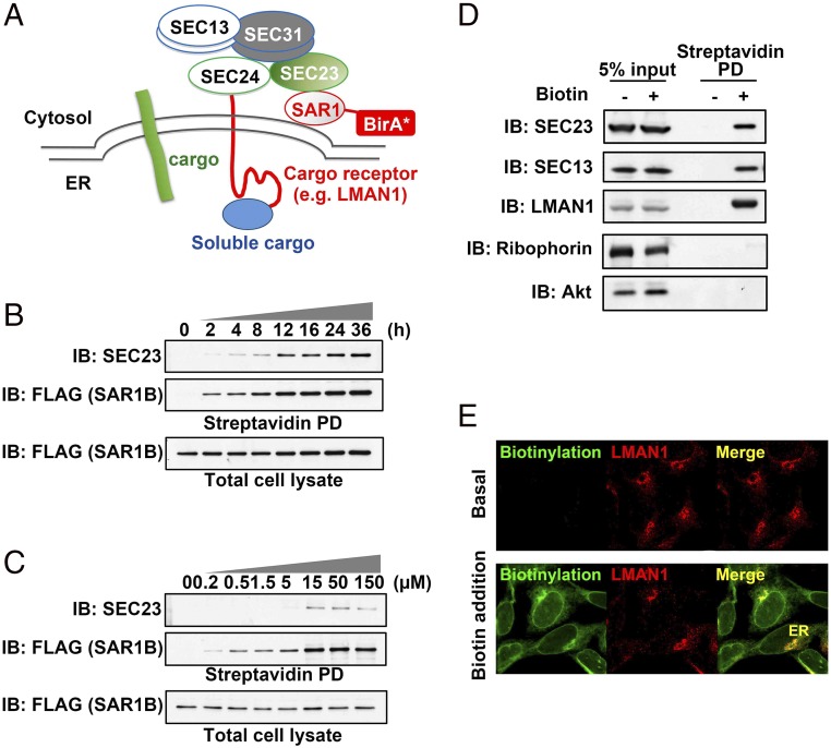
Fig. 1.
Development of a proximity-dependent biotinylation assay for COPII-mediated cargo transport. (A) Overall scheme of proximity-dependent biotinylation of the COPII machinery using SAR1-BirA*. Activated SAR1 initiates the assembly of the COPII coat, which recruits cargos and/or additional regulatory factors (e.g., the cargo receptor LMAN1) to defined microdomains, allowing biotinylation by BirA* fused to SAR1. (B) Time course of biotinylation of the COPII subunit SEC23 by SAR1B-BirA*. 293A cells stably expressing SAR1B-BirA* (with a FLAG tag at the C terminus of BirA*) were treated with 15 µM biotin for different time points as indicated. After cell lysis, biotinylated proteins were isolated by streptavidin bead pull-down (PD) and subjected to SDS/PAGE followed by immunoblotting (IB) with anti-SEC23 (Top) or anti-FLAG (recognizing SAR1B; Middle). Immunoblotting for SAR1B-FLAG in total cell lysates is shown (Bottom). The anti-SEC23 antibody recognizes both SEC23A and SEC23B. (C) Biotin dose dependence of SEC23 labeling by SAR1B-BirA*. 293A cells stably expressing SAR1B-BirA* were treated for 4 h with different doses of biotin as indicated. After cell lysis, biotinylated proteins were isolated by streptavidin beads and subjected to SDS/PAGE followed by immunoblotting with the indicated antibodies. (D) Capture of COPII cargos by SAR1B-BirA* in vivo. 293A cells stably expressing SAR1-BirA* were treated with 15 µM biotin for 4 h. After cell lysis, biotinylated proteins were isolated by streptavidin beads and subjected to SDS/PAGE followed by immunoblotting with the indicated antibodies; “5% input” indicates total cell lysate equivalent to 5% of the material used for streptavidin pull-down in lanes 3 and 4 (“Streptavidin PD”). (E) Subcellular localization of biotinylated proteins. 293A cells stably expressing SAR1B-BirA* were treated with 15 µM biotin for 4 h. After cell fixation, biotinylated proteins were visualized by immunostaining and confocal microscopy with an anti-LMAN1 antibody or Alexa Fluor-conjugated streptavidin.

