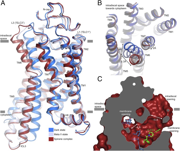
Fig. 4.
A ligand channel in S-RS1–stabilized rhodopsin. (A) Side view and (B) top view onto the superposition of the S-RS1–bound conformation (red) of opsin-, dark- (PDB ID: 1GZM, blue), and meta-II–state rhodopsin (PDB ID: 3PQR, blue-white) visualizing the helical movements in TM6 and TM7 at the intradiscal side of the membrane. The complex has a higher structural similarity to meta-II (rmsd = 0.4 Å) than dark-state rhodopsin (rmsd = 1.6 Å). (C) Ligand channel in surface representation sliced perpendicular to the membrane highlighting an intradiscal opening formed between L2 (“ECL2”), TM5, and TM6a as well as a membrane opening, large enough to permit the exchange of hydrophobic ligands including retinal.

