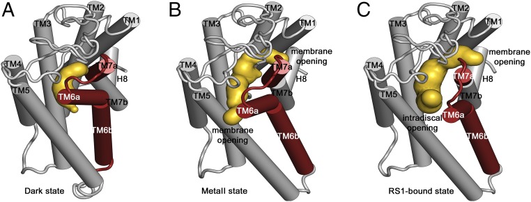
Fig. 5.
Ligand channel comparison of dark, meta-II, and S-RS1–bound opsin. Cartoon representation of the receptor depicting unaltered transmembrane helix regions (gray) and region of ligand-induced conformational changes (red). The caver analysis reveals the void pathways of the receptor in yellow of (A) dark-state rhodopsin bound to 11-cis retinal featuring an occluded pocket, (B) light-activated meta-II rhodopsin with bound all-trans retinal showing two small openings toward the membrane, and (C) S-RS1–bound opsin identifying a ligand channel toward the membrane and intradiscal space suggesting a potential route for retinal exchange and ligand-induced receptor breathing.

