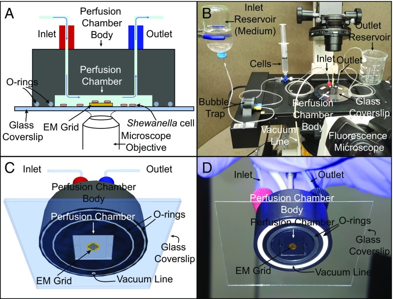
Fig. 1.
Schematic and actual images of the perfusion flow imaging platform (objects not drawn to scale). (A and B) Cross-sectional (A) and 3D (B) views of the perfusion flow imaging platform. An electron microscopy (EM) grid is glued to a glass coverslip that seals the perfusion chamber. S. oneidensis cells injected into the sealed chamber attach to the grid surface and are sustained by a continuous flow of the medium. Cells are labeled with the fluorescent membrane dye FM 4-64FX and monitored in real time for OM extension growth using an inverted fluorescent microscope placed under the perfusion chamber. (C and D) A 3D schematic (C) and image (D) of the perfusion chamber interior with an attached EM grid.

