Fig. 5.
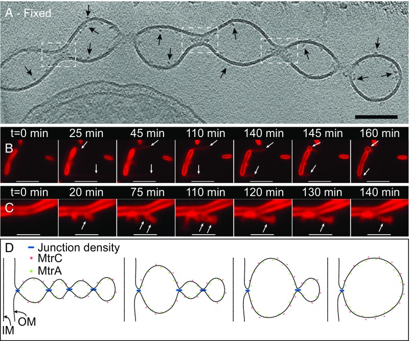
Proposed model for the formation and stabilization of OMV chains. (A) ECT image of a chemically fixed OM extension reveals the presence of densities at junctions that connect one vesicle to the next along the OMV chain (white dashed boxes). While all of the junction densities are not visible in the tomographic slice in A, Movie S6 is a 3D reconstruction of the same OM extension revealing the densities present at every junction. In addition, densities possibly related to decaheme cytochromes can be observed on the interior and exterior of the OM along the extension (arrows). (Scale bar: 100 nm.) (Fig. S8.) (B and C) Time-lapse fluorescence images recorded in real time in the perfusion flow imaging platform monitoring the growth and transformation of an OM extension from an apparently long filament (OMV chain morphology) to a single large vesicle (B, indicated by arrows) in S. oneidensis Δflg (a mutant strain lacking flagellin genes). (Movie S7.) Movie S8 shows OM extensions from wild-type cells also exhibiting a similar behavior to Δflg and a large vesicular morphology to an apparently smoother filament (OMV chain morphology) (C, indicated by arrows) in wild-type S. oneidensis MR-1 cells. (Movies S9 and S10). The cells and the OM extensions in B and C are stained by the membrane stain FM 4-64FX. (Scale bars in B and C: 5 µm and 2 µm, respectively.) (D) Schematic depicting a hypothesis for the formation and stabilization mechanism of OMV chains: Junction densities on the interior of the OM extension facilitate the constriction of the membrane, enabling the formation of an OMV chain. These constriction densities can be removed or added to facilitate transformation of an OMV chain to a large vesicle or vice versa as observed in B and C, respectively.

