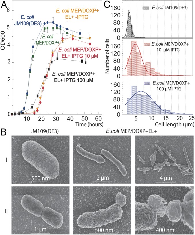
Fig. 3.
Growth and cell morphology analysis of the heterochiral mixed membrane strains. (A) Growth of the E. coli MEP/DOXP+EL+ strain with all of the ether lipid enzymes [not induced (orange)], induced with 10 μM (red), and induced with 100 μM (black) of IPTG added early during growth (OD600 = 0.0) compared with two negative control strains: E. coli JM109(DE3) wild-type (blue) and E. coli MEP/DOXP+ strain with the integrated MEP-DOXP operon (green). The data are the averages of three biological replicates ±SEM. (B) SEM of wild-type E. coli and the heterochiral mixed membrane strain induced at a late (0.3 OD600) and early (0.03 OD600) growth phase using 100 μM of IPTG. (I) Altered cell shape and length. (II) aberrant cell division and formation of bulges and shreds. (C) Statistical analysis of the cell length of the E. coli JM109(DE3) (Top), E. coli MEP/DOXP+EL+ induced with 10 μM IPTG (Middle), and 100 μM IPTG (Bottom).

