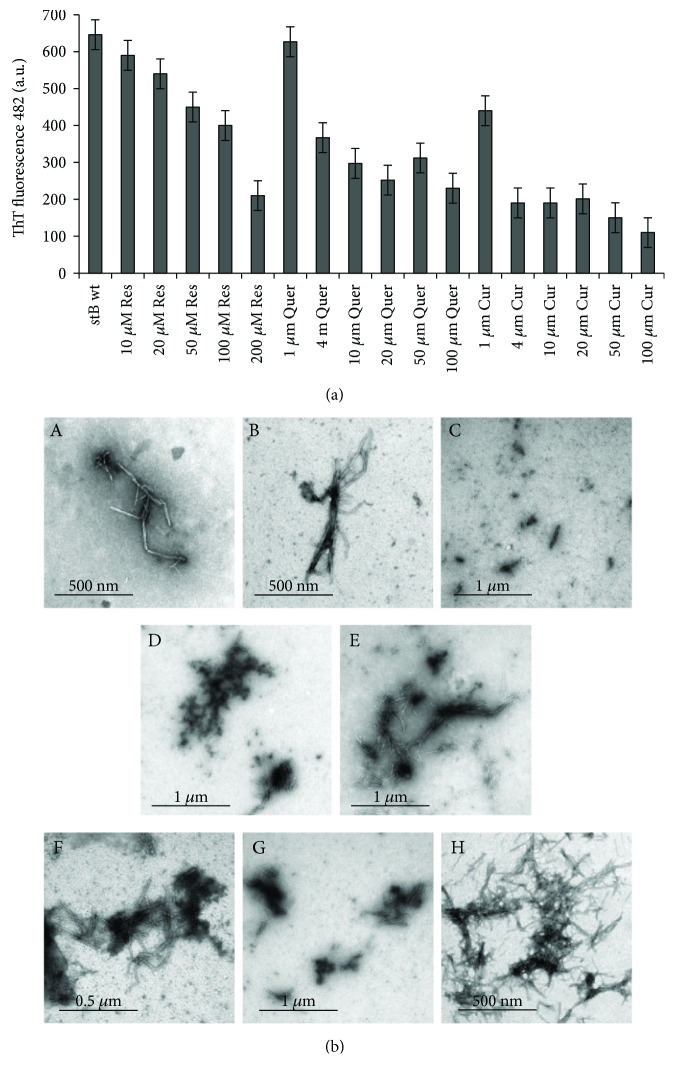
Figure 9.
ThT fluorescence intensity versus TEM data. (a) Graph of ThT fluorescence intensity in the plateau phase of amyloid fibrillation versus antioxidant concentration. (b) TEM data. Concentration dependence of fibril morphology and approximate amounts; samples were taken in the plateau phase of the reactions of amyloid fibril formation by stB at different polyphenol concentrations: (A) 1 μM Cur, (B) 10 μM Cur, (C) 50 μM Cur, (D) 1 μM Quer, (E) 50 μM Quer, (F) 50 μM Res, (G) 100 μM Res, and (H) 200 μM Res.

