Figure 1.
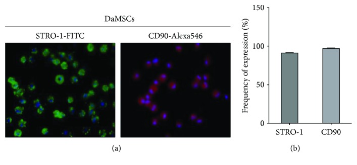
Immunolocalization of mesenchymal stem cell makers in deer antler-derived mesenchymal stem cells (DaMSCs). Immunocytochemistry staining was conducted on DaMSCs with primary antibodies directed against STRO-1 (a, left, green), and CD90 (a, right, red) and stained by FITC- and Alexa 546-conjugated secondary antibodies, respectively. Nuclei were visualized with DAPI (blue). (b) Graph indicates quantification of protein expression. Data represent the mean ± s.d. (n = 3).

