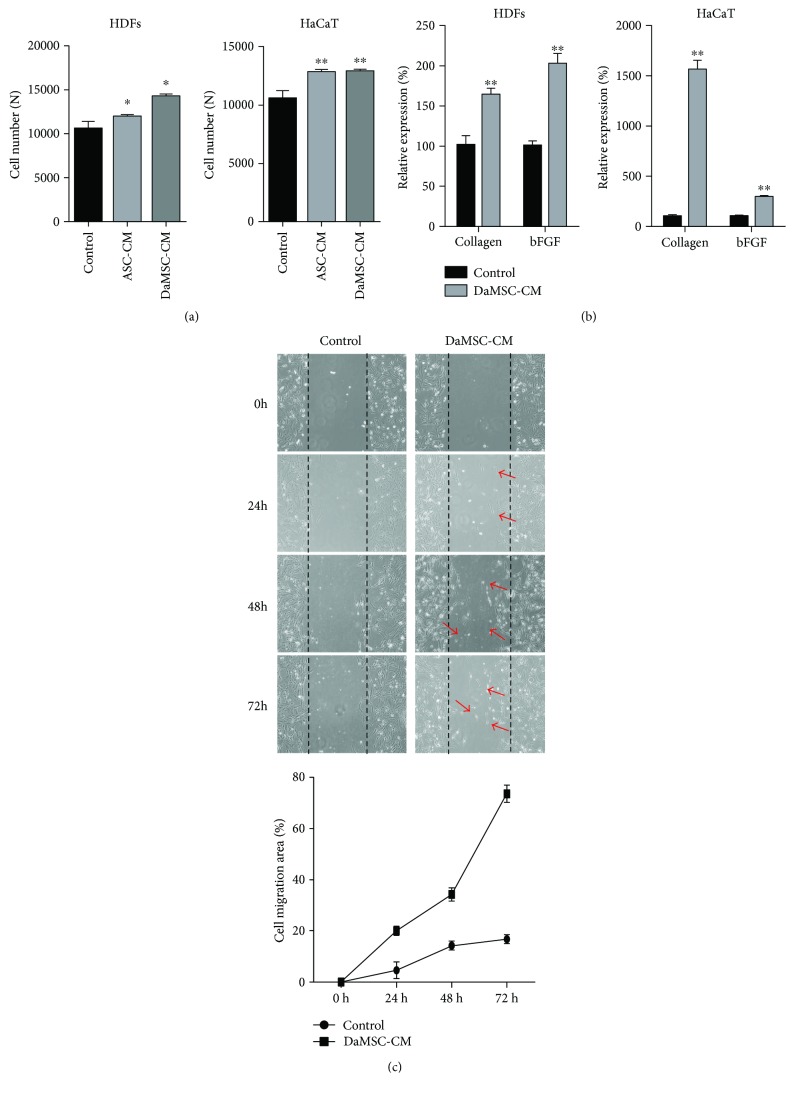
Figure 4.
Effects of skin regeneration on treatment with DaMSC-CM. (a) Proliferation of human dermal fibroblasts (HDFs) and keratinocytes (HaCaTs) under treatment of three different types of media for 48 h. (b) qRT-PCR analysis showing relative expression of collagen type Ι mRNA (b, left) and basic fibroblast growth factor (bFGF) mRNA (b, right), normalized to expression of GAPDH mRNA. (c) In vitro wound healing assay exhibiting HDF migration upon treatment with DaMSC-CM in a time-dependent manner. Red arrows indicate cell migration. Black lines represent wound spaces created with plastic micropipette tips. Data represent the mean ± s.d. (n = 3). ∗ P < 0.01, ∗∗ P < 0.001 by one-way analysis of variance with Tukey's multiple comparison tests, as compared with the control.

