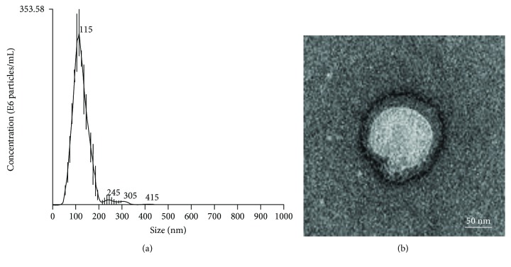
Figure 5.
Characterization of extracellular vesicles (EVs) derived from DaMSCs. (a) Nanoparticle tracking analysis (NTA) showed a size distribution of EVs with an average of 119.9 nm. (b) Transmission electron microscopic (TEM) images revealed the morphology of the EVs with a membrane bilayer. Scale bar indicates 100 nm. Particle number: 2.29 × 10¹⁵ particles/mL.

