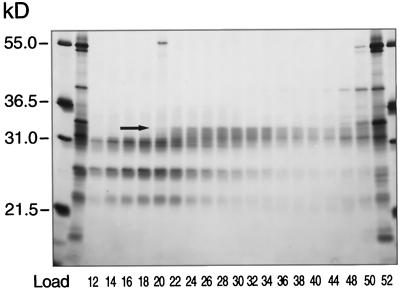
Figure 4.
SDS-PAGE of the crystalline HA column fractions. Proteins present in fractions from the crystalline HA column were separated by SDS-PAGE. Electrophoresis was carried out on a 20- × 20-cm gel containing a 10% to 13% acrylamide gradient, and the gel was stained with silver. The arrow indicates a protein band at 33 kD whose staining intensity in the various fractions correlates with WS activity detected in those same fractions.

