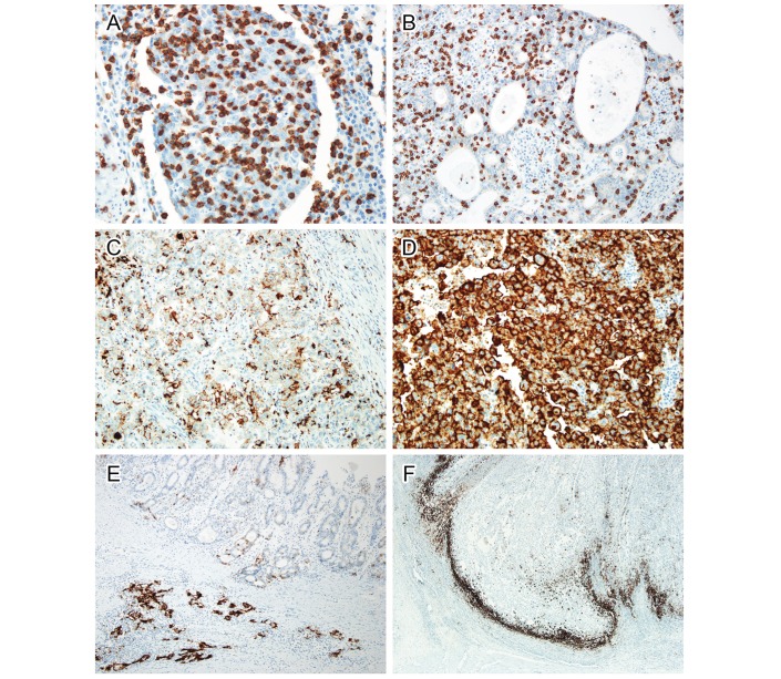
Figure 1.
Immunohistochemistry of CD8 (A and B) and PD-L1 (C–F). Intratumoural CD8-positive T cells were more (A) or less (B) numerous than cancer cells. In PD-L1 staining, tumour cells were weakly stained in (C) or strongly stained (D) in the cellular membrane. Both intensities were considered positive staining. In some cases, tumour cells demonstrated marked heterogeneity in PD-L1 expression (E). Well-differentiated tumour cells on the surface were PD-L1 negative, while infiltrating cell nests in the submucosal layer were strongly positive for PD-L1. PD-L1 was predominantly stained in immune cells at the invasive front (F). PD-L1, programmed death-ligand 1.

