Figure 1.
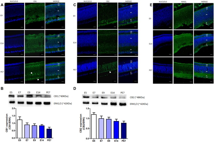
Expression of selective endocannabinoid markers during avian retina development. Expression of CB1, CB2 and monoacylglycerol lipase (MAGL) using immunofluorescence and western blotting. In (A), expression of CB1 (green) at embryonic stages E9 and E14 and post-hatched 7 days (PE7) in chick retina. At PE7, CB1 is mainly found in cells of the inner nuclear layer (INL) and ganglion cell layer (GCL; white arrows). (B) Western blot highlights CB1 expression from embryonic day 5 (E5) to PE7. Expression of ERK 1/2 was used as loading control. In (C), CB2 (green) expression from E9 to PE7 primarily in the inner plexiform layer (IPL) lamina (white arrow). CB2 expression from E5, as shown in (D). In (E), MAGL expression (green) from E9 to PE7 is shown, its distribution is evident across all layers of the retina. Scale bar of 20 μm, all images were obtained with 40× magnification in a Zeiss fluorescence microscope. N = 6 for each analysis.

