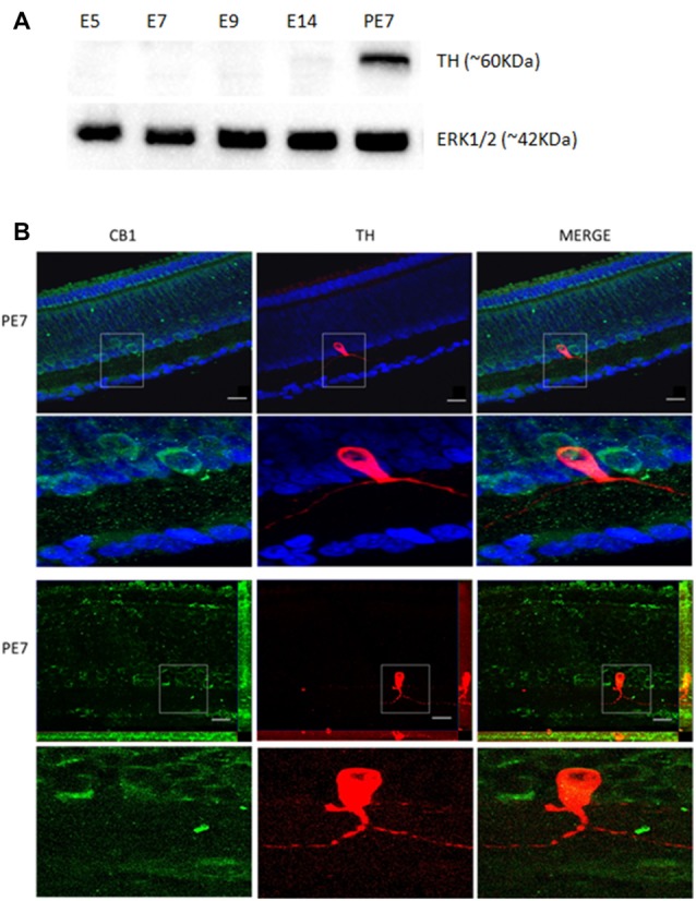
Figure 2.

Co-localization of CB1 receptor and tyrosine hydroxylase (TH) enzyme in the avian retina. We evaluated whether dopaminergic neurons, TH positive cells (red) express CB1 receptor (green) by immunofluorescence. In (A), western blotting for TH at different stages, confirming that TH is highly expressed at PE7. ERK1/2 is shown as the loading control. In (B), CB1 and TH co-expression in PE7 avian retina. As shown, a discrete TH co-expression with CB1 that is mainly present in neighboring cells in the INL. ApoTome images in upper row and confocal images in lower row. Scale bar of 20 μm, all images were obtained with 40× magnification. N = 5 for each analysis.
