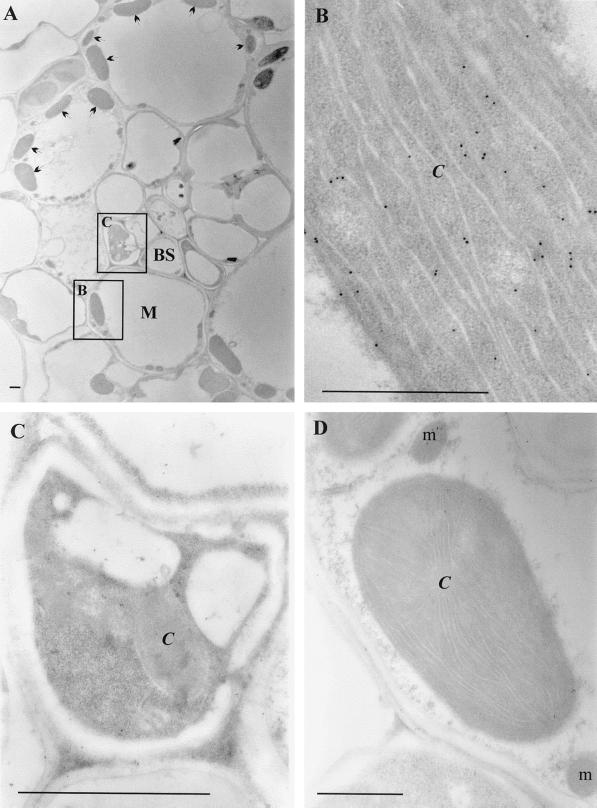
Figure 2.
Immunogold localization of GR in maize leaves. A to C, Sections incubated with GR antibody (1:100 dilution). D, Sections incubated with preimmune serum control. C, Chloroplast; BS, bundle sheath cells; M, mesophyll cells; and m, mitochondrion. Arrows indicate the position of chloroplasts in the mesophyll cells. Squares in A indicate the types of tissues examined in B and C. Bars = 1 μm.

