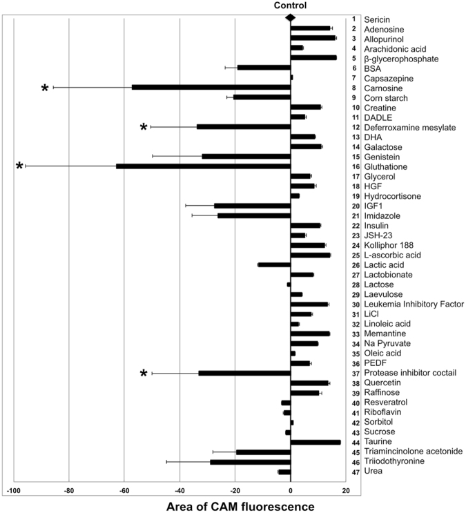
Figure 1.
Cell viability after ten days of storage as measured by area of CAM fluorescence. The control line drawn from the black diamond represents the area of calcein-acetoxymethyl ester (CAM) fluorescence obtained in the control group (N = 18), where 1% sericin was added to the MEM-based storage medium. Other bar points are representations of CAM area fluorescence for each additive (N = 3) supplemented to MEM in the presence of 1% sericin. Resulting effects are displayed as either increasing or decreasing CAM area fluorescence compared to the control line. The addition of carnosine, deferroxamine mesylate, glutathione or the protease inhibitor cocktail to the storage medium significantly reduces cell viability as measured by CAM area fluorescence (*, P < 0.05). Error bars represent the standard deviation of mean values. BSA: bovine serum albumin; DADLE: [D-Ala2, D-Leu5]-Enkephalin; DHA: docosahexaenoic acid; HGF: hepatocyte growth factor; IGF1: insulin-like growth factor 1; JSH-23: 4-methyl-1-N-(3-phenylpropyl)benzene-1,2-diamine; PEDF: pigment epithelium-derived factor.

