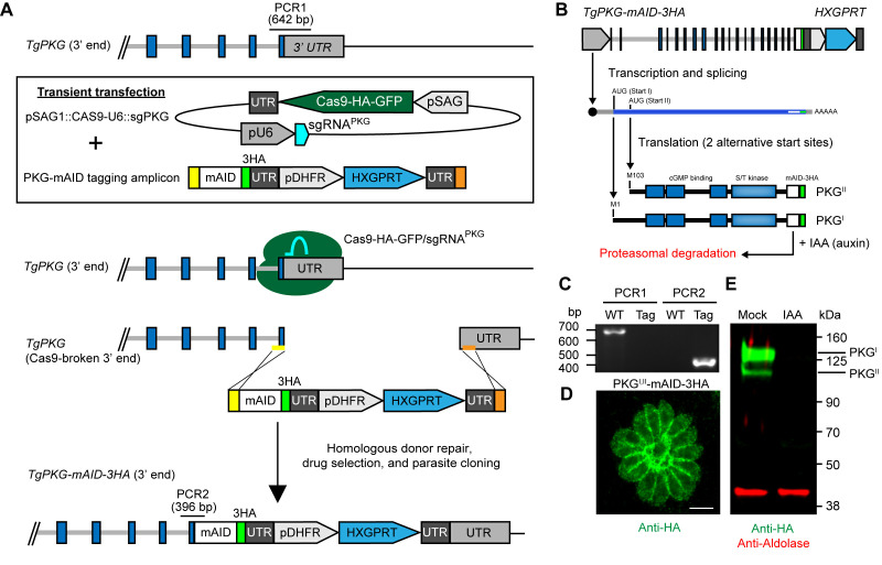
Figure 3. Generation and regulation of (m)AID protein fusions.
A. CRISPR-mediated C-terminal mAID tagging in RH TIR1-3FLAG line. The example shown here is the gene encoding protein kinase G (TgPKG, reproduced with permission from [ Brown et al., 2017 ]). Note the locations of diagnostic PCRs 1 and 2. B. Schematic of TgPKG-mAID-3HA expression showing the two PKG-mAID isoforms that are generated from alternative translation initiation sites on the transcript. Addition of IAA promotes the simultaneous degradation of both PKG-mAID isoforms. C-D. Data reproduced with modification for space limits from ( Brown et al., 2017 ). C. Diagnostic PCRs 1 and 2 from genomic DNA samples showing successful integration of the mAID tagging construct into TgPKG (see A for PCR positions). WT (wild type), TIR1-3FLAG parent; Tag, PKGI,II-mAID-3HA parasites. D. Immunofluorescence of PKG-mAID-3HA isoforms I and II stained with anti-HA mAb and detected with Alexa Fluor 488-conjugated secondary antibody. Scale bar = 5 µm. E. Western blot assay of lysed PKGI,II-mAID-3HA parasites probed with antibodies recognizing HA (green) and aldolase (red). Parasites were treated with 500 µM IAA or the vehicle (EtOH) for 4 h prior to lysis.

