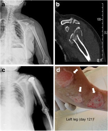
Fig. 1.

Radiograpic findings of the left shoulder and ruptrured blisters in the left leg. a: Plain radiograph showing the patient’s left shoulder at admission. b: CT scans of the same area. c: Plain radiograph of the left shoulder after hemiarthroplasty surgery. d: During the treatment failure stage, the patient developed multiple blisters in the left leg. Defects in the skin developed after the blisters ruptured (arrows)
