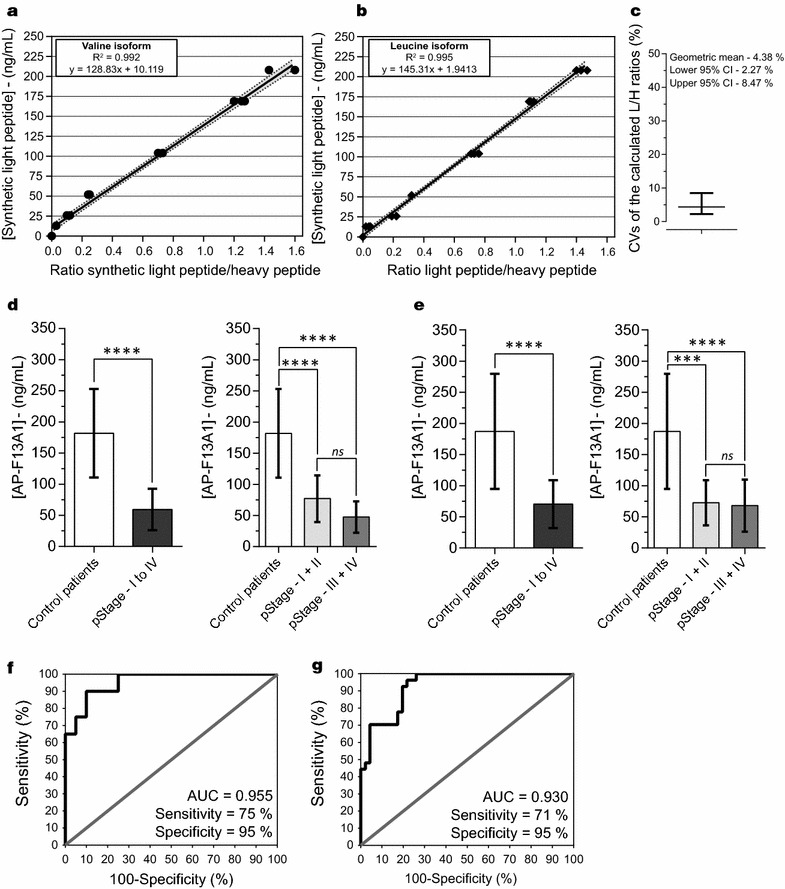
Fig. 3.

Calibration curves and evaluation of AP-F13A1 fragments by LC-PRM assays. a, b Graphical plot representation of theoretical concentration spanning according to the experimental peak ratio light/heavy peptides for AVPPNNSNAAEDDLPTVELQGVVPR and AVPPNNSNAAEDDLPTVELQGLVPR. c Coefficients of variations (CVs) of the light/heavy ratios distribution for the two peptides, over the 6 concentration points. Serum concentration of AP-F13A1 in healthy patients and patients with declared CRC from the first sample bank (d) and the second sample bank (e). The calculated means were 181.8 ± 71.0 and 187.2 ± 92.2 ng/mL for the controls, and 59.4 ± 33.3 and 70.4 ± 38.4 ng/mL for the cancer patients, both showing significant Wilcoxon tests (****P value = 0.0001). Receiver Operating Characteristic curves (ROC) for AP-F13A1 from first sample bank (f) and the second one (g). AUC computing were 0.95 (0.89–1.00) and 0.93 (0.87–0.98) and calculated values of sensitivity/specificity were 75%/90% and 71%/95%, respectively
