
Figure 13.
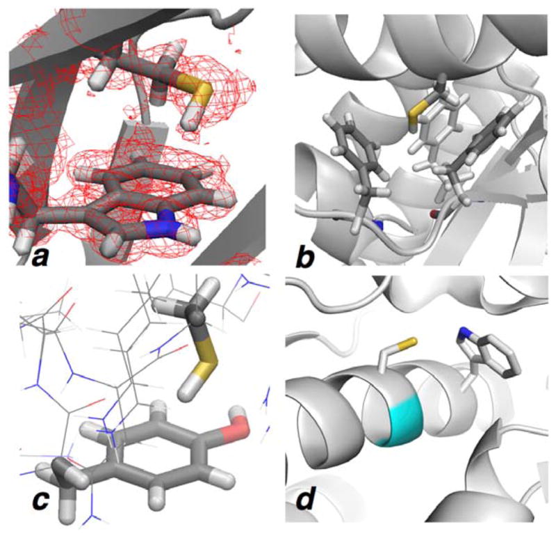
S–H/π interactions in the PDB. (a) Cys79-Trp40 S–H/π interaction in pdb 3puc (0.96 Å resolution), with a difference electron density map (coefficients 2Fo–Fc) shown, that exhibits alignment of the S–H bond with the aromatic π orbital.32 (b) Cys306 interacting in a cage of 3 phenylalanine residues in pdb 3u3h (0.97 Å resolution). The electron density map is unclear about the hydrogen position, though no traditional hydrogen bond acceptor is near the thiol.33 (c) Cys87-Phe181 S–H/π interaction (2.63 Å H...Caromatic distance) in pdb 4txr (1.00 Å resolution).34 Notably, all S–H bonds in the pdb files of (a)–(c) included non-canonical 1.20 Å S–H bond lengths and 109.0˚ C–S–H bond angles. (d) W216XXGC220 interaction within the F-helix of the protein kinase CK2 (pdb 3war, 1.04 Å resolution).36 Gly219 is indicated in cyan.