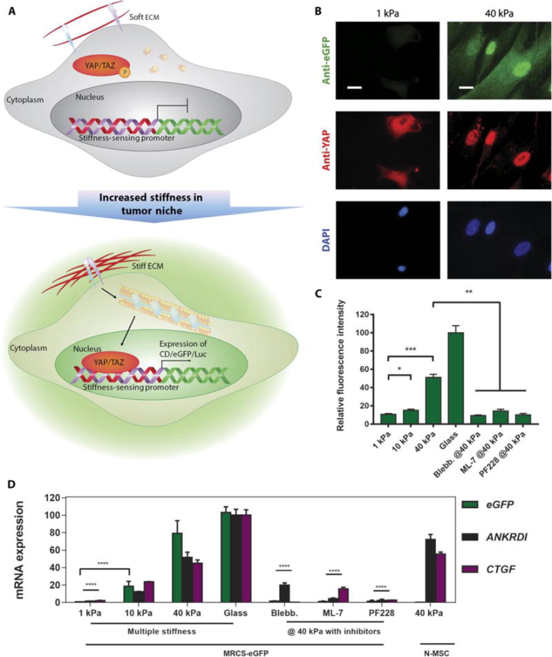
Fig. 1. MRCS in vitro validation.

(A) Schematic of a proposed mechanism of how MRCS works. When the stiffness of ECM increases, YAP/TAZ are activated and localize to the nucleus. Then, YAP/TAZ will bind to the synthetic stiffness-sensing promoter in MRCS and drive the expression of downstream reporters (such as eGFP and Luc) and/or therapeutics. Note: This schematic is simplified to clarify the major components in MRCS mechanism. (B) Representative images of MRCS-eGFP plated on soft (~1 kPa) and firm (~40 kPa) polyacrylamide gels. eGFP (stained with anti-eGFP; green) was turned on in response to higher stiffness. YAP (stained with anti-YAP; red) localization is also regulated by stiffness, such that it concentrates in the nuclei on stiffer substrates. 4′,6-diamidino-2-phenylindole (DAPI) (blue; nuclear counterstain) is displayed. Scale bars, 25 μm. (C) Quantification of fluorescence intensity of eGFP (stained with antibody) from MRCS-eGFP seeded on substrates with different stiffness or on firm (~40 kPa) substrates treated with 10 μM ML-7 (MLCK inhibitor) or 20 μM PF228 (FAK inhibitor). Blebb., blebbistatin. Data aremeans ± SEM. (D) RT-qPCR analysis of MRCS-eGFP on hydrogels. Expression of eGFP (green) and YAP/TAZ downstream factors (CTGF, purple; ANKRDI, black) was increased on stiffsubstrate and was down-regulated on soft substrate or with mechanosensing inhibitors, showing that MRCS is stiffness-specific. Quadruplicate samples were used for the analysis. Data are means ± SD. *P < 0.05, **P < 0.01, ***P < 0.001, and ****P < 0.0001.
