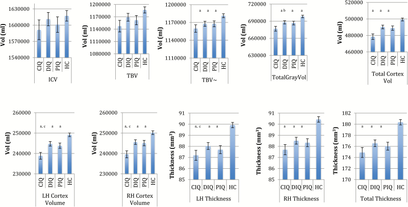
Fig. 1.
Mean differences in global brain measures that survived false discovery rate (FDR) correction across healthy controls (HC), compromised patients (CIQ), deteriorated patients (DIQ), and preserved patients (PIQ). LH, left hemisphere; RH, right hemisphere; Vol, volume; ~TBV co-varying for ICV; error bars represent standard errors. Global volume analyses adjusted for ICV, age, gender, and site. Thickness analyses adjusted for age, gender, and site. aDifferent to HC (P < .05 FDR corrected). bCIQ different to DIQ only (P < .05 FDR corrected). cCIQ different to DIQ and PIQ (P < .05 FDR corrected).

