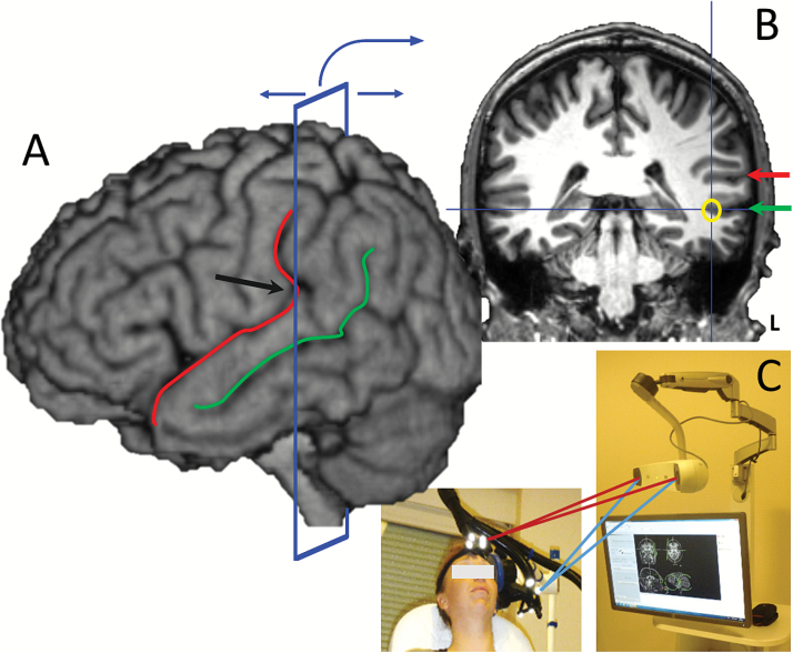
Fig. 2.
Location of the rTMS stimulation target by structural MRI and neuronavigation (A) Before rTMS treatment, the lateral sulcus (red line) and the superior temporal sulcus (green line) are located on the 3D MRI view using neuronavigation software. Then the coronal plane is moved in the posterior direction until it crosses the point where the lateral sulcus becomes vertical (black arrow). (B) The lateral sulcus (red arrow) and the superior temporal sulcus (green arrow) are located based on the 3D view on the coronal plane. Finally, the rTMS target is defined at the bottom of the superior temporal sulcus (yellow circle). (C) The neuronavigation tool is used during treatment to precisely locate the coil over the target.

