
Figure 2.
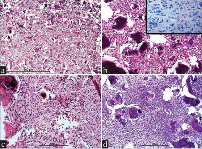
Case 2 (a and b) and Case 3 (c and d) showing basophilic psammomatoid ossicles in a cellular background of fibroblastic tissue. Tumor cells are negative for epithelial membrane antigen (inset, B) (a = H and E, ×100; b = H and E, ×400, c and d = H and E, ×200)