


Figure 2.
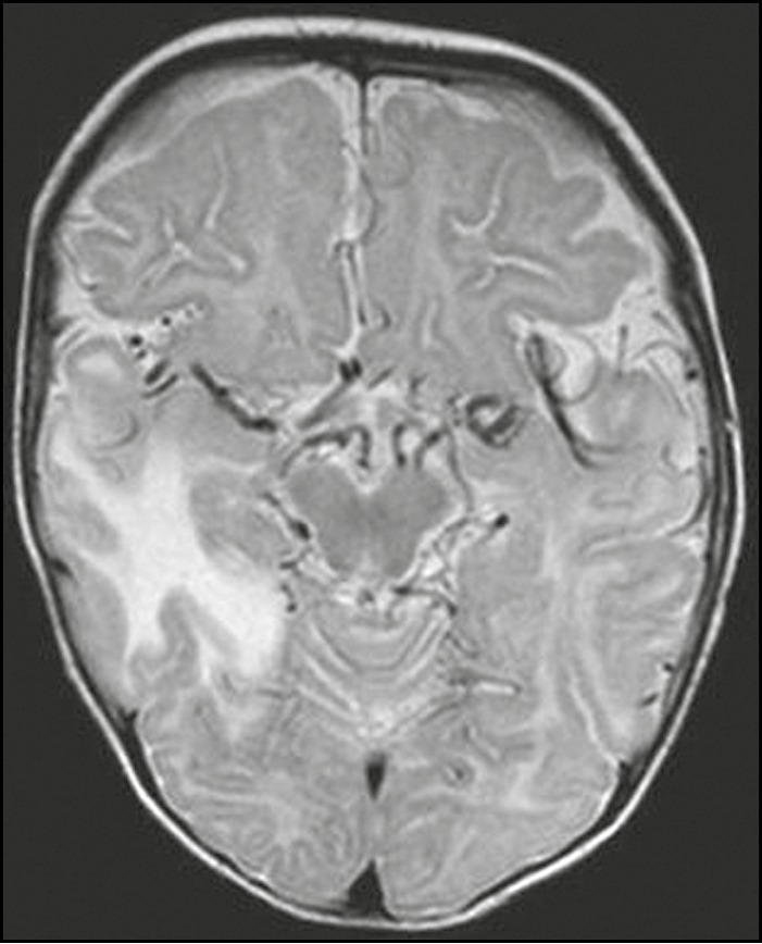
Axial T2-weighted image highlighting marked tortuosity of vessels within the basal cisterns. The child also had swollen right temporal lobe, a recognized feature of Menkes disease
Official websites use .gov
A
.gov website belongs to an official
government organization in the United States.
Secure .gov websites use HTTPS
A lock (
) or https:// means you've safely
connected to the .gov website. Share sensitive
information only on official, secure websites.
Axial T2-weighted image highlighting marked tortuosity of vessels within the basal cisterns. The child also had swollen right temporal lobe, a recognized feature of Menkes disease