
Fig. 2.
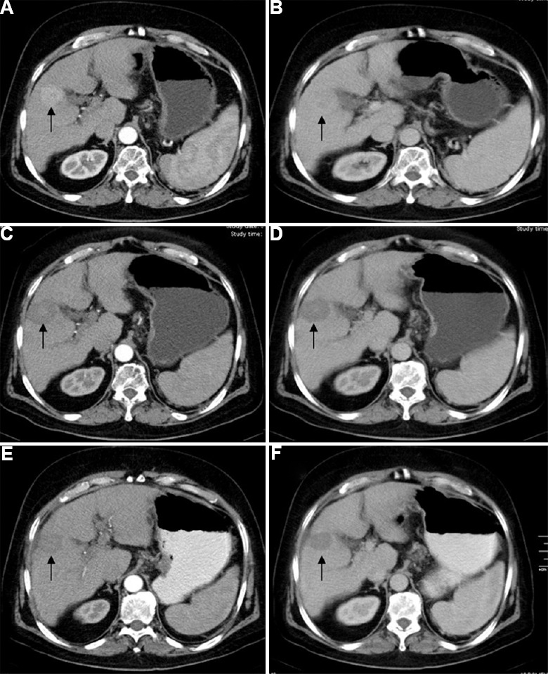
A 65 yr old male with hepatocellular carcinoma in segment 5 of liver treated with radiofrequency ablation alone. Pre-treatment arterial (A) and venous (B) phase images showing hypervascular lesion in segment 5 of liver (arrows). Post-radiofrequency ablation follow up imaging at three months (C & D) and six months (E & F) shows no residual enhancement in the lesion (arrows).