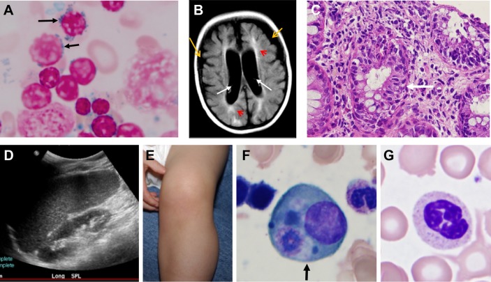
Figure 2.
Clinical findings of the NHGRI cohort of patients with SIFD. (A) Iron staining of patient 2 bone marrow aspirate showing ring sideroblasts (that represented more than 50% of erythroid precursors) (black arrows) and increased iron staining. (B) Abnormal findings in the cerebrum of patient 3 by fluid-attenuated inversion recovery (FLAIR) MRI of the brain. The image shows bilateral cerebral atrophy (yellow arrows), enlarged ventricles (white arrows) and extensive leukomalacia (red arrow heads). (C) H&E staining of rectosigmoid colon biopsy in patient 3 showing acute focal inflammation. (D) Splenomegaly in patient 2. (E) Knee effusion in patient 5. (F) Phagocyte (arrow) on bone marrow aspirate smear of patient 4. (G) Neutrophil with toxic granules in peripheral blood smear of patient 4. SIFD, sideroblastic anaemia with immunodeficiency, fevers and developmental delay.

