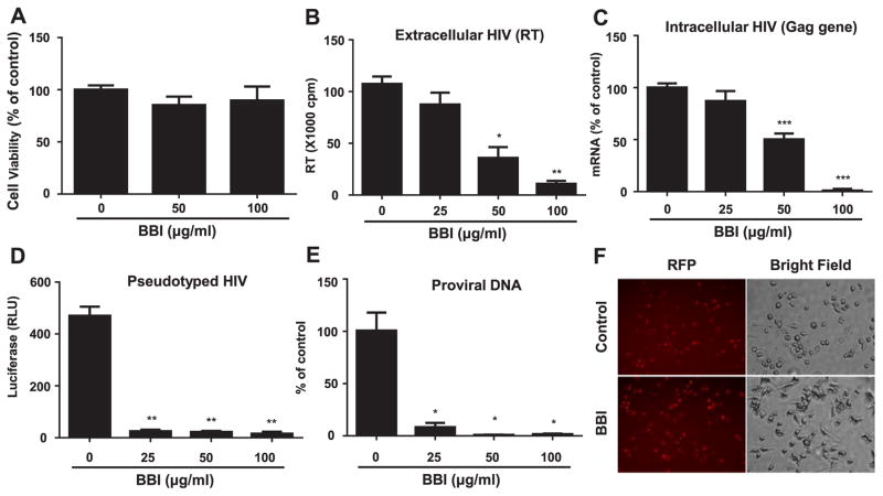
Fig. 1. BBI inhibits HIV infection of macrophages at the entry level.
(A) Seven-day-cultured macrophages were treated with/without BBI at indicated concentrations for 6 days. The cell viability was assessed by MTS assay. The data are expressed as the absorbance (490 nm) relative to untreated control, which is defined as 100%. Data are shown as mean±SD for three independent experiments. Macrophages derived from monocytes of the healthy donors were treated with/without indicated concentrations of BBI for 24 h prior to HIV (Jago) infection. After washing away unbound virus, fresh medium without BBI was added to the cultures. Cells and cell-free supernatant were collected on day 5 post-infection for HIV reverse transcriptase (RT) assay (B). Data are shown as mean±SD for three independent experiments. (C) Cellular RNA was subjected to real time RT PCR for HIV Gag and GAPDH RNA. The data are expressed as HIV RNA levels relative (%) to untreated control, which is defined as 100%. Data are shown as mean±SD for three independent experiments. (D) Macrophages were treated with/without the indicated concentrations of BBI for 24 h prior to pseudotyped HIVADA infection. After washing away unattached virus, fresh medium without BBI was added. Macrophages were cultured for additional 48 h and then lysed with cell lysis buffer. Luciferase activities (relative light units, RLU) were measured. The data are expressed as the RLU change vs untreated control. Data are shown as mean±SD for three independent experiments. (E) Macrophages were treated with/without the indicated concentrations of BBI for 24 h prior to HIV (Jago) infection. Macrophages were cultured for additional 24 h and then DNA was extracted from the cells. The proviral DNA was quantified with the real time PCR. Data are shown as mean±SD for three independent experiments. (*P<0.05, **P<0.01, ***P<0.001 when performing Student’s t-test). (F) Macrophages were treated with/without BBI (100 μg/ml) for 24 h prior to VSV-G pseudotyped HIV infection for additional 24 h. The RFP protein expression was observed under a fluorescent microscope.

