Fig. 3.
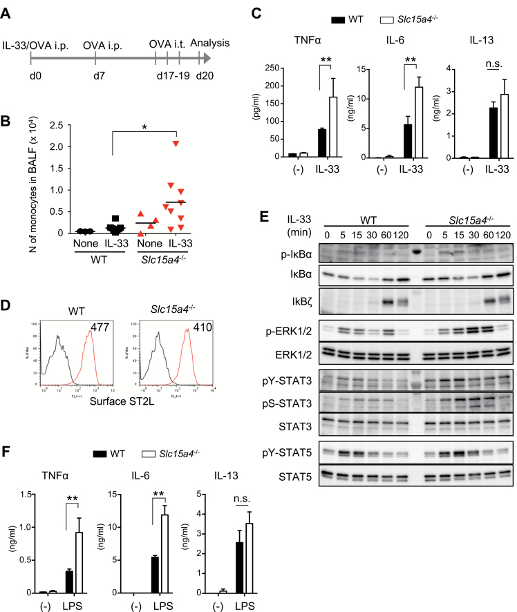
SLC15A4-deficiency alters IL-33 signaling in mast cells. (A) Schematic of the IL-33-induced airway inflammation model. (B) Number of monocytes in the BALF before or after IL-33-induced inflammation in WT or Slc15a4−/− mice, analyzed by flow cytometry. Bars indicate the mean, and data are integrated from three independent experiments (n = 3 for WT none, n = 8 for WT IL-33, n = 4 for Slc15a4−/− none, n = 9 for Slc15a4−/− IL-33). *P < 0.05. (C) Cytokine production in response to IL-33 stimulation in BMMCs. TNFα, IL-6 and IL-13 levels in culture supernatants of WT or Slc15a4−/− BMMCs stimulated with recombinant IL-33 for 6 h measured by ELISA (in technical triplicate). Results are representative of four independent experiments. **P < 0.01; n.s., not significant. (D) Surface expression of the IL-33 receptor ST2L on BMMCs, analyzed by flow cytometry. (E) IL-33 signaling in WT or Slc15a4−/− BMMCs was analyzed by immunoblotting. WT or Slc15a4−/− BMMCs stimulated with recombinant IL-33 for the indicated periods were subjected to immunoblot analysis with the specific antibodies. (F) Cytokine production in WT or Slc15a4−/− BMMCs in response to LPS stimulation for 6 h; cytokine levels in the culture supernatant were measured by ELISA (in technical triplicate). For (D–F), results are representative of three independent experiments. **P < 0.01; n.s., not significant.

