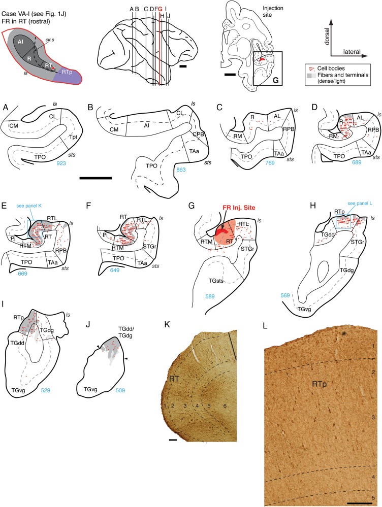
Figure 10.
Connections of area RT in case VA-l. (A–J) Distribution of both retro- and anterograde labeling in the STP, STG, and temporal pole areas after FR injection into the rostral part of area RT (section G; see also Fig. 1J). No label was observed in AI, caudal belt, or Tpt (A and B) and only a few filled cells were found in caudal R (C). Cells and terminals were observed in rostral R (D), near the border with RT. Label was most dense within RT caudal to the injection site (E, F, and K). Labeled cells and fibers were dense in RTL and RTM (E and F), but sparse in RPB (D and E). Label rostral to the injection was restricted to area RTp (H, I, and panel L) and the dorsal temporal pole (J). Scale bars = 5 mm in (A–) and 0.2 mm in (K and L). For other conventions see Figure 4.

