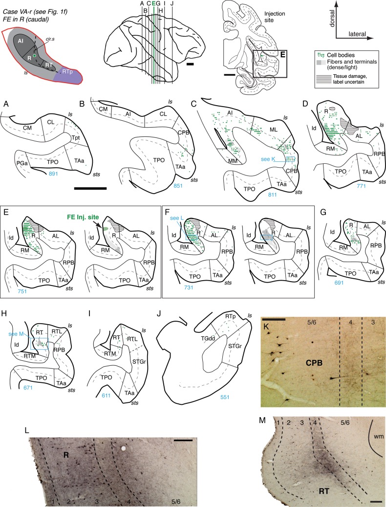
Figure 7.
Connections of area R in case VA-r. (A–J) Distribution of retrograde and anterograde label in the auditory-related areas after FE injection into the caudal portion of area R (section E; see also Fig. 1F). No cells or terminals were found in caudal core or belt (A and B). Labeled cells and patches of anterograde label were located in rostral AI, ML, and CPB (C and K). The strongest label was located within area R (D–F and L); for clarity, retrograde and anterograde label are displayed separately in panels E and F. Relatively few cells were found rostral to the injection site, predominantly within R (G). In RT, anterograde label was concentrated in layer 4 and deep layer 3 of central RT (section H and panel M). Only a few cells were found in RT and RTp (H–J). Scale bars = 5 mm in panels A–J, and 0.2 mm in K–M. For other conventions see Figure 4.

