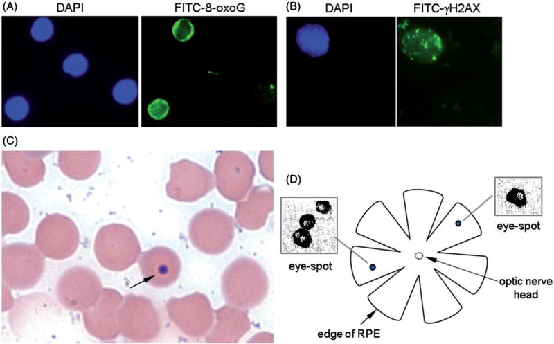
Figure 1.
Genotoxicity assays used. (A) Immunofluorescence of 8-oxoG. Cellular nuclei are shown by DAPI staining and 8-oxoG is visualized by FITC staining. (B) Immunofluorescence of γ-H2AX. Cellular nucleus is visualized by DAPI staining and γ-H2AX foci are visualized by FITC staining. (C) Micronucleus assay. A Giemsa-stained peripheral blood smear with a micronucleated erythrocyte highlighted by an arrow is shown. (D) DNA deletion assay. Schematic of RPE and close-up of eye-spots are shown.

