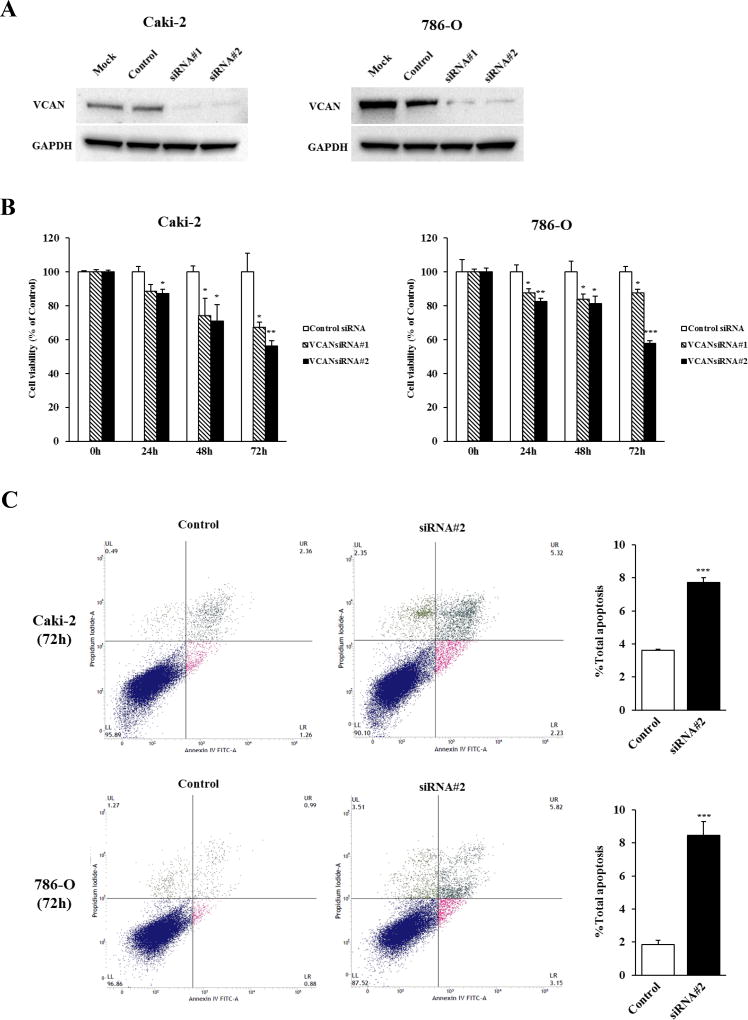
Figure 2. Effect of VCAN knockdown on cell proliferation and apoptosis in ccRCC cells.
(A) Knockdown of VCAN in Caki-2 and 786-O cells was determined by immunoblot analysis at 72 hours after transfection with two different VCAN siRNAs (#1 and #2). GAPDH used as loading control. (B) Cell viability was analyzed by the MTS cell proliferation assay at 0, 24, 48 and 72 hours after treatment with siRNAs. The attenuation of VCAN significantly inhibited cell viability in a time-dependent manner in both cell lines. *, P<0.05. **, P<0.01. ***, P<0.001. (C) Apoptosis assays with Caki-2 and 786-O cells were done 72 hours post siRNA#2 transfection by flow cytometric analyses. Left: Representative biparametric histograms showing cell population in early apoptotic (bottom right quadrant), apoptotic (top right quadrant), and viable (bottom left quadrant) states for each treatment. Right: Bar chart indicates the total apoptotic cell fraction percent (early plus apoptotic) in VCAN siRNA#2 transfectants compared with controls. ***, P<0.001.

