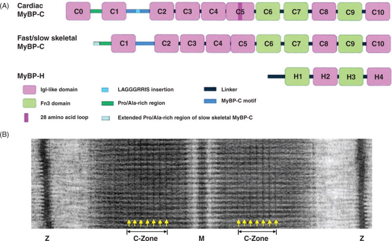
Figure 15.

Myosin-binding proteins (MyBP). (A) Schematic drawing of MyBP domain organization. MyBPs are composed of a series of immunoglobulin (Igl-like in pink) and fibronectin type III (Fn3 in green) repeat domains. Domains termed C1 through C10 and a 105-residue linker between C1 and C2 termed the MyBP-C motif (in blue) make up the core structure of MyBP-C isoforms. Cardiac MyBP-C has the addition of an eight IgI-like domain termed C0, a unique amino acid sequence—LAGGGRRIS—insertion (in light blue) in the MyBP-C motif, and a 28 amino acid insertion (in dark pink) in the C5 domain. Slow skeletal MyBP-C differs from the fast isoform with an extended Pro/Ala-rich region at the N-terminus. MyBP-H is the smallest isoform with four domains similar to C7 through C10 of MyBP-C and a unique Pro/Ala-rich linker (in black) region. (B) Example electron micrograph of frog skeletal muscle showing MyBP-C transverse stripes located in the C-Zone. [Part A modified, with permission, from (167); Part B modified, with permission, from (406).]
