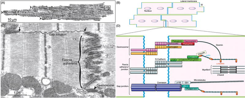
Figure 18.

Structural organization and molecular components of the intercalated disc (ICD). Low-magnification transmission electron micrograph (A) and schematic drawing of cardiac myocardium (B) exhibit characteristic step-like structures of intercalated discs (A, arrowheads) formed through syncytial interconnection of rod-shaped cardiomyocytes. (C and D) Higher magnification view of areas enclosed in A and B, respectively, show three specialized substructures of intercalated discs—fascia adherens (adherens junction), desmosome (desmosomal junction, arrowhead), and gap junction. The ends of gap junction here connect to two adherens junctions (arrows). (D) Molecular components of ICD substructures not only serve as mechanical and electrochemical coupling platforms between adjacent cardiomyocytes, but also interact with major cytoskeletal filament systems (e.g., actin and microtubule cytoskeletons). The following proteins are not discussed in texts: p120, p150, EB1, and protein 4.3). [Parts A and C reprinted, with permission, from (626); B and D reprinted, with permission, from (38).]
