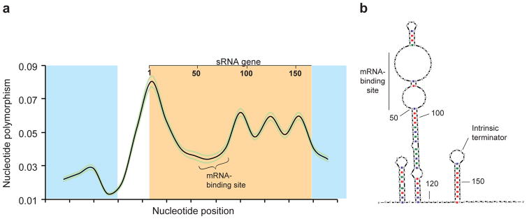
Figure 2. Sequence conservation and structure of an sRNA gene.
(a) Sequence conservation within an sRNA gene (orange) and flanking protein-coding genes (blue). The black line represents nucleotide diversity index, π, calculated using a sliding window analysis; the flanking green lines indicate the 95% confidence interval. Lowest nucleotide polymorphism within sRNA genes is observed in mRNA-binding regions. (b) Predicted structure of an sRNA, showing a single-stranded mRNA-binding site and a terminator hairpin. Adapted from Kacharia et al. 2017.

