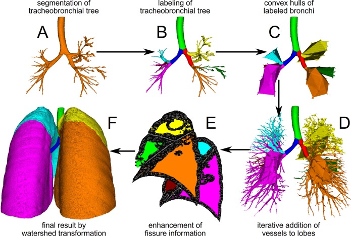
Fig 1. Work-flow chart for fully automatic lung lobe segmentation.
A: The initial step is the segmentation of the airway tree. B: Second, central airways and lobar bronchi are labeled by an anatomical knowledge-based algorithm. C: Then, a convex hull around the labeled lobar bronchi is generated. D: In the next step vasculature is iteratively subsequently segmented as far into the lung periphery as possible and added to the corresponding lobe by distance measures. E: Lastly, fissures are detected by eigenvalue/eigenvector operations (sagittal view right and left lung). F: Final results of automatic lobe segmentation using bronchi, vessel and fissure information as volume rendering images in posterior view. Lobes are indicated as follows: yellow = right superior lobe, green = middle lobe, orange = right inferior lobe, light blue = left superior lobe, red = lingual, pink = left inferior lobe.

