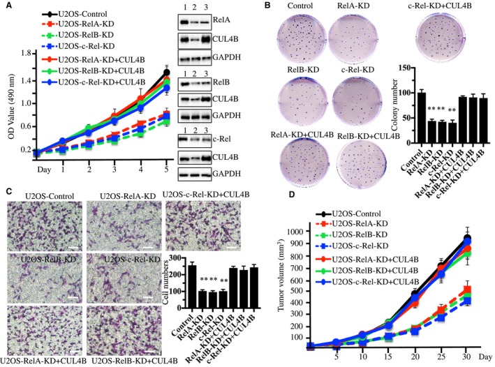
Figure 3.

Knockdown of RelA, RelB, or c‐Rel inhibits osteosarcoma cell growth. (A) Knocking down RelA, RelB, or c‐Rel inhibited osteosarcoma cell proliferation. U2OS cells were transfected with Con‐shRNA (U2OS‐Control), RelA‐shRNA (U2OS‐RelA‐KD), RelB‐shRNA (U2OS‐RelB‐KD), c‐Rel‐shRNA (U2OS‐c‐Rel‐KD), RelA‐shRNA + pCDNA3‐CUL4B (U2OS‐RelA‐KD + CUL4B), RelB‐shRNA + pCDNA3‐CUL4B (U2OS‐RelB‐KD + CUL4B), or c‐Rel‐shRNA + pCDNA3‐CUL4B (U2OS‐c‐Rel‐KD + CUL4B). After a 48‐h incubation, MTT assays were conducted to evaluate cell proliferation with absorbance measurement at 490 nm. The knockdown efficiency of RelA, RelB, and c‐Rel was measured with western blots (right panel). 1 (U2OS‐Control), 2 (U2OS‐RelA, RelB, or c‐Rel‐KD), 3 (U2OS‐RelA, RelB, or c‐Rel‐KD + CUL4B). (B) Knocking down RelA, RelB, and c‐Rel decreased colony formation rates. Cells used in A were seeded onto 12‐well plates and cultured with 0.1 mL of fresh medium containing 0.5% FBS for two weeks. **P < 0.001 (C) Knocking down RelA, RelB, and c‐Rel decreased cell invasion. Cells used in A were subjected to Boyden chamber assays, and the invasive cells were counted (right panel). **P < 0.001. Bars=100 μm. (D) Knocking down RelA, RelB, and c‐Rel decreased the in vivo tumor formation ability. Cells used in A were injected intradermally into the flanks of mice. Tumor volumes were measured with fine calipers at 5‐day intervals.
