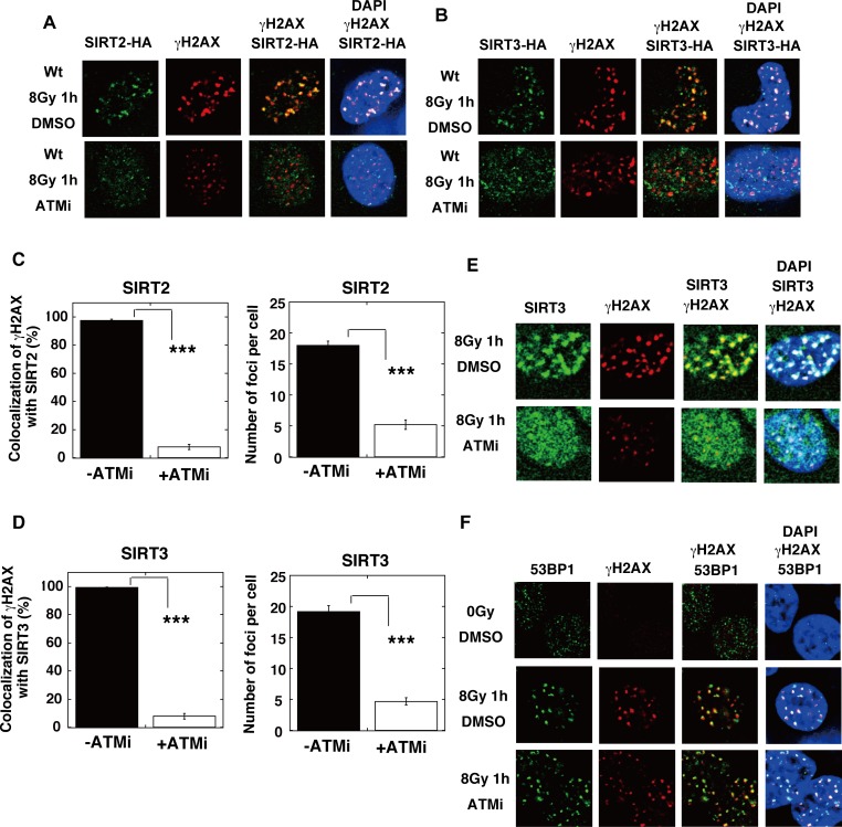
Fig 13. ATM inhibition impedes the recruitment of SIRT2/SIRT3, but not 53BP1, at IR-induced DSB sites.
(A, B) T-Rex-293 cells expressing SIRT2-HA (A), or SIRT3-HA (B) were irradiated with γ-rays (8 Gy). The KU55933 (ATMi) solution or the same volume of DMSO was added to the cells 1 h before irradiation. At 1 h after irradiation, the cells were subjected to immunofluorescent staining. Immunofluorescent images with an anti-HA (green) antibody, an anti-γH2AX (red) antibody, and DAPI (blue). (C, D) The percentages of γH2AX foci colocalization with SIRT2 (C) or SIRT3 (D) in Fig 13A and 13B were calculated, as described in the Supporting Materials and Methods, and are shown in the graphs. The numbers of protein foci per cell are also shown in the graphs. Error bars indicate the standard error of the mean. Asterisks indicate statistically significant differences between the indicated pairs of groups (***, p<0.001 by t-test). (E, F) The KU55933 solution or the same volume of DMSO was added to T-Rex-293 cells, 1 h before irradiation with γ-rays (8 Gy). (E) Cells were subjected to immunofluorescent staining at 1 h after irradiation with an anti-SIRT3 (green) antibody, an anti-γH2AX (red) antibody, and DAPI (blue). (F) Cells were subjected to immunofluorescent staining at 1 h after irradiation with an anti-53BP1 (green) antibody, an anti-γH2AX (red) antibody, and DAPI (blue). As a control, unirradiated cells (0 Gy) were used.

