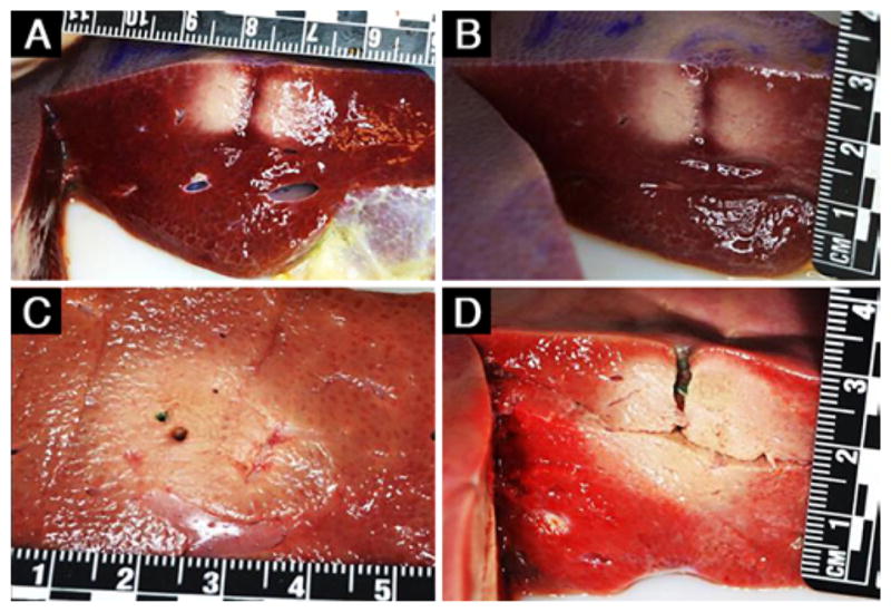
FIG. 4.

Representative gross pathology images of the ablated pig liver tissue obtained with the single transducer element (A and B) and 2-element (C and D) configurations, each with a single insertion of the Acoustx applicator. The rulers are marked in centimeters. The image in D was obtained after TTC staining.
