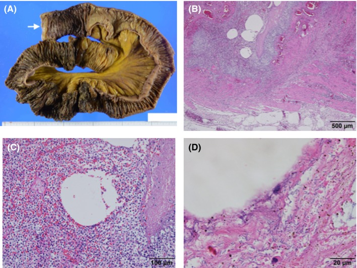
Figure 3.

Histological observation of the resected small intestine of an 84‐year‐old man with necrotizing enterocolitis associated with Clostridium butyricum, using hematoxylin–eosin staining with a macro image. A, Macro image of resected tract. Arrow indicates the oral side. B, Severe inflammation, several vacuoles, congestion, and hemorrhage in the muscularis propria and subserosal layer indicate gas gangrene. Scale bar, 500 μm. C, Vacuoles are surrounded by acute severe inflammation and considered as gas accumulation. Scale bar, 100 μm. D, Observation by oil immersion lens reveals numerous bacilli adjacent to vacuoles. Scale bar, 20 μm.
