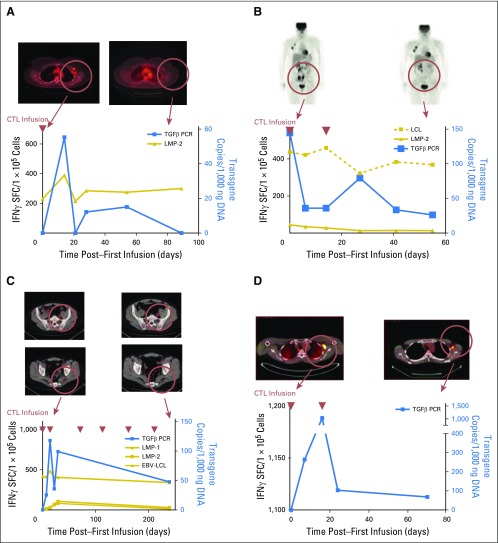
Fig 3.
Clinical responses and immune reconstitution. (A) In patient 2, whose axillary lymph nodes responded to an infusion of dominant-negative transforming growth factor-β (TGF-β) type 2 receptor (DNRII) latent membrane protein (LMP)–specific T cells (DNRII-LSTs; as shown by positron emission tomography [PET]/computed tomography scan), there is a rise in both DNRII-transduced T cells as detected by quantitative PCR and LMP-2–specific T cells as detected by interferon-gamma (IFN-γ) enzyme-linked immunospot (ELISPOT) assay. At the time the patient achieved a complete remission, 2 months postinfusion, DNRII-transduced T cells were no longer detectable in the peripheral blood. (B) A PET scan demonstrating increased uptake in the ilium is observed in patient 3. The follow-up scan 8 weeks post–T-cell infusion 1 shows a partial response that coincided with an increase in DNRII-transduced T cells by quantitative PCR and detectable Epstein-Barr virus (EBV)–specific T cells (against lymphoblastoid cell line targets) and, to a lesser extent, LMP-2–specific T cells (against LMP-2 pepmix) by IFNγ ELISPOT assay. (C) A PET scan demonstrating increased uptake in the lumbar spine and sacrum is observed in patient 5 after receiving both radiotherapy and donor lymphocyte infusion post–allogeneic stem-cell transplatation. The follow-up scan 8 weeks post–donor-derived DNRII-LST infusion 1 shows a good partial response that coincided with an increase in DNRII-transduced T cells by quantitative PCR and EBV-, LMP-1–, and LMP-2–specific T cells as determined by IFNγ ELISPOT assay. (D) A PET scan demonstrating increased uptake in the axilla is observed in patient 8. The follow-up scan 8 weeks post–DNRII-LST infusion 1 shows overall stable disease and an increase in DNRII-transduced T cells by quantitative PCR. Blue lines denote quantitative PCR copies of the DNRII transgene, gold lines denote spot-forming cells as measured by IFNγ ELISPOT assay, and red arrows denote days of T-cell infusions. SFC, spot-forming cell.

