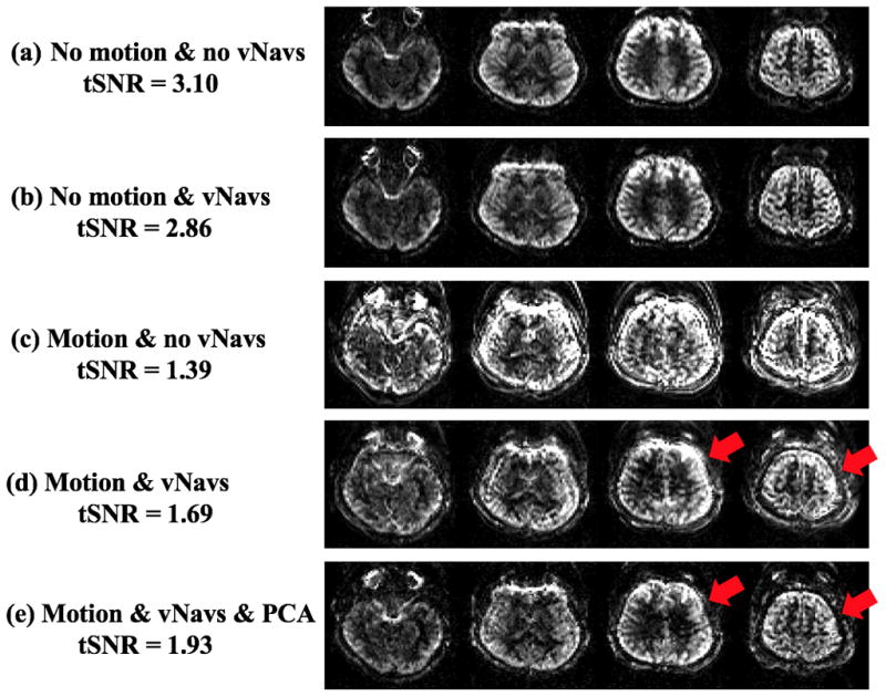
Figure 3.

Four representative slices of perfusion weighted signal. (c) and (d) were acquired when the volunteer was prompted to move once a minute. (b) and (d) was motion corrected by vNavs during real-time acquisition. (e) was corrected with PCA off-line and showed the same data as in (d).
