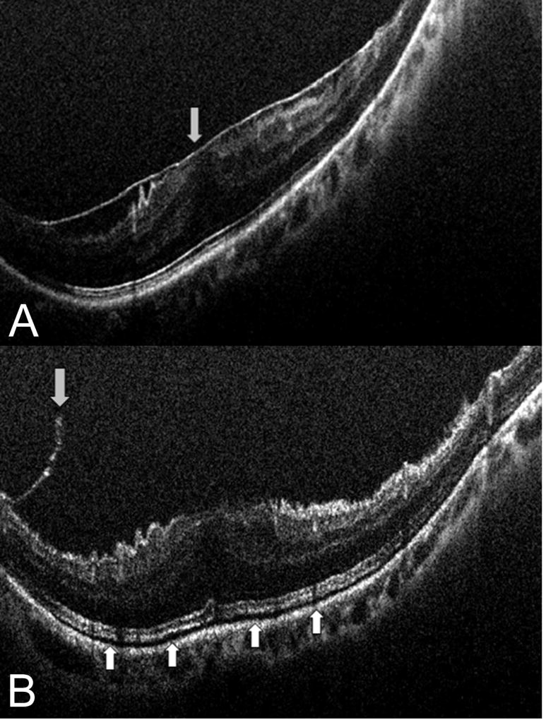
Figure 1. Intraoperative optical coherence tomography and epiretinal membrane peeling.
(A) Intraoperative optical coherence tomography (OCT) prior to membrane peeling with clearly identified epiretinal membrane. (B) Post-peel intraoperative OCT with occult residual membrane (down arrow) and increased subretinal hyporeflectance (up arrows).

