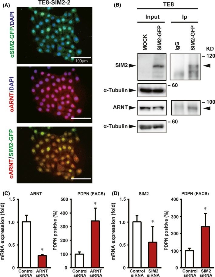
Figure 6.

SIM2 and ARNT cooperatively decrease PDPN‐positive tumor basal cells. A, Immunofluorescence showing colocalization of SIM2‐GFP and ARNT in the nuclei of TE8‐SIM2‐2 cells. B, Immunoprecipitation of SIM2‐GFP with anti‐green fluorescent protein (GFP) antibody 1 day after transient SIM2‐GFP transfection to TE8 cells followed by western blot with anti‐SIM2, ‐ ARNT, or‐α tubulin antibody. Input, unprecipitated extracts. IgG, control IP. C, Real‐time RT‐PCR of ARNT (left) and percentage of PDPN‐positive cells (right) in KYSE510 cells that were transfected with control siRNA (white column) or ARNT siRNA (red column) (n = 3, mean + SE; *P < .05). D, Real‐time RT‐PCR of SIM2 (left) and percentage of PDPN‐positive cells (right) in KYSE510 that were transfected with control siRNA (white column) or SIM2 siRNA (red column) (n = 3, mean + SE; *P < .05)
