Figure 4.

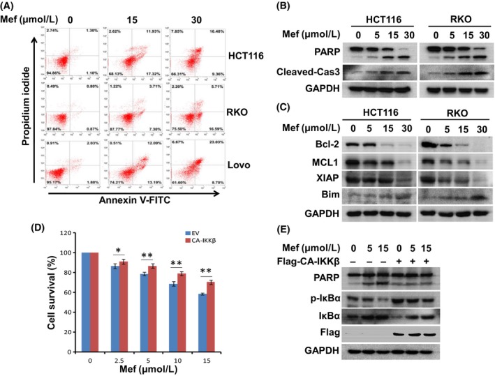
Mefloquine (Mef) induces colorectal cancer cell apoptosis. A, HCT116, RKO and Lovo cells were treated with mefloquine for 12 h and then stained with Annexin V‐FITC and propidium iodide. Percentages of annexin V+ are indicated in the scatterplot (right low and upper quadrants). B, HCT116 and RKO cells were incubated with 0, 5, 15, 30 μmol/L mefloquine overnight and then evaluated by immunoblotting against PARP, cleaved‐caspase 3 and GAPDH. C, HCT116 and RKO cells were treated with 0, 5, 15, 30 μmol/L mefloquine overnight and then analyzed by immunoblotting against anti‐apoptotic (Bcl‐2, Mcl1 and XIAP) and pro‐apoptotic (Bim) proteins. GAPDH was used as a loading control. D, Empty vector (EV) or CA‐IKKβ plasmids were transfected into HCT116 cells. Cells were treated with indicated concentrations of mefloquine overnight and then evaluated by CCK‐8 assay. IKK, IκB kinase. *P < .05; **P < .01, as compared with control. E, Empty vector (EV) or CA‐IKKβ plasmids were transfected into HCT116 cells. The cells were treated with indicated concentrations of mefloquine overnight and then evaluated by immunoblotting against PARP, p‐IκBα, IκBα, Flag and GAPDH
