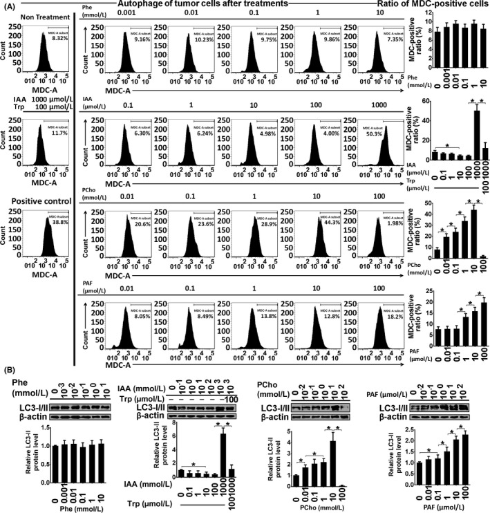
Figure 7.

Autophagy assessment by monodansylcadaverine (MDC) staining (A) and LC3‐I/II immunoblotting (B) in Ishikawa cells treated with phenylalanine (Phe), indoleacrylic acid (IAA), phosphocholine (PCho) and platelet‐activating factor‐16 (PAF) for 48 hours. *P < .05
